Newly Found T. Rex Blood Vessels Give A Rare Glimpse Into Dinosaur Biology
Some are quick to point out that movies like Jurassic Park may get the appearance of dinosaurs wrong. If so, it is for good reason. Scientists primarily have fossils of bones and teeth to guide their understanding of how dinosaurs appeared. Soft tissue, such as muscle or fat, has long been lost to the passage of time. That piece means we are missing a major part of how dinosaurs looked, lived, and how their biology functioned overall.
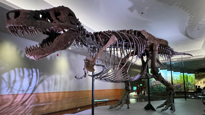
A recent study utilized a Tyrannosaurus rex fossil — though not the newly discovered T. rex fossil covered in armor – that was at the Royal Saskatchewan Museum in Canada. The study was published in Scientific Reports under the title "In situ analysis of vascular structures in fractured Tyrannosaurus rex rib". It brought together five researchers from the University of Regina in Canada, and it may have just opened up the door for revolutionary soft tissue research.
From a partially healed bone injury, the research team was able to use a series of advanced imaging techniques to study where the T. rex would have bled and how the injury healed. Their findings provide scientists with a rare glimpse into the internal processes that occurred inside a living dinosaur. This opportunity presents a new pathway for future fossil studies, and it can broaden our understanding of dinosaur soft tissue as a whole.
What was discovered inside the T. rex fossil
The T. rex fossil in the study had a fractured rib that drew the researchers' attention, knowing that this mostly healed injury could give some insight into dinosaur soft tissue. The research team faced two major challenges. The first, of course, was to ensure that no damage was done to this important fossil during the evaluation process. The second was that they were working with a fossil, not a fresh bone, which limited the amount of analysis that could be done.
First, the team sliced tiny sections of bone and evaluated them under microscopes to see how the bone was built and how it had changed over time. Then they used X-ray scans to create detailed 3D images of the inside of the bone, which revealed blood vessel-like shapes. To determine the composition of these structures, researchers mapped the chemical elements in the fossil using advanced scanning techniques. Finally, they zoomed in with an electron microscope to get an even closer look at the bone's makeup.
They discovered that the blood vessel structures they found are mineral casts, primarily composed of iron minerals such as pyrite and rust. These likely formed from a mix of minerals in the surrounding soil and potentially iron from the dinosaur's own blood. The rib had been fractured and was starting to heal when the T. rex died. Although we know where dinosaurs died based on their fossils, research is making headway into understanding where they originated in the first place.
What this means for the future of dinosaur biology research
Looking to the future, the research team suggests that examining more fossils with signs of injuries or disease could reveal more about dinosaur biology, including how their bones healed. By comparing blood vessel structures across different species, including living creatures like birds and crocodiles, scientists can build a more comprehensive picture of dinosaur health, growth, and evolution.
The approach employed in this study presents a promising avenue for future research. The techniques utilized here can be applied to help find more preserved soft tissue structures. It can also help expand our understanding of the medical challenges of creatures that lived millions of years ago, such as the new research regarding dinosaur cancer.
We may no longer have the original soft tissue to discover and analyze dinosaurs, but their secrets can still be uncovered in the fossilized bones. It will be exciting to follow future studies that build upon the work done here. Perhaps one day, we'll know exactly how dinosaurs truly looked.